

Graduate Student at Rice
Mary (Lingbo) Jin
Hi, my name is Mary and I am a 5th-year graduate student at Rice Computational Imaging Lab advised by Professor Ashok Veeraraghavan. My research interest is in leveraging computational imaging for seeing below the skin applications. Specifically, I’m interested in jointly designing the optical system and post-processing algorithm to improve the resolution and contrast of clinical microscopy. My projects are done in collaboration with Dr.Yubo Tang from Rice Optical Spectroscopy & Imaging Lab.
Education & Experience
Rice University
Graduate Student
Department of ECE
2018 – Present
Intuitive Surgical
applied research intern
Summer 2021
Genentech
Intern
Summer 2018
Carnegie Mellon University
Bachelor of Science
Department of ECE
Department of Biomedical Engineering
2014-2018
Recent Projects
2019-2020
Deep learning extended depth-of-field microscope for fast and slide-free histology
Traditional microscopy suffers from a fixed trade-off between depth-of-field (DOF) and spatial resolution—the higher the desired spatial resolution, the narrower the DOF. We present DeepDOF, a computational microscope that allows us to break free from this constraint and achieve >5× larger DOF while retaining cellular-resolution imaging—obviating the need for z-scanning and significantly reducing the time needed for imaging. The key ingredients that allow this advance are 1) an optimized phase mask placed at the microscope aperture; and 2) a deep-learning-based algorithm that turns sensor data into high-resolution, large-DOF images. DeepDOF offers an inexpensive means for fast and slide-free histology, suited for improving tissue sampling during intraoperative assessment and in resource-constrained settings.
Lingbo Jin, Yubo Tang, Yicheng Wu, Jackson B. Coole, Melody T. Tan, Xuan Zhao, Hawraa Badaoui et al. “Deep learning extended depth-of-field microscope for fast and slide-free histology.” Proceedings of the National Academy of Sciences (2020).

The DeepDOF image clearly visualizes subcellular features, primarily individual nuclei that are evenly distributed on the esophageal wall. Multiple ROIs are shown in regions where the conventional microscope images are out of focus (red) and in focus (cyan).
2018-2019
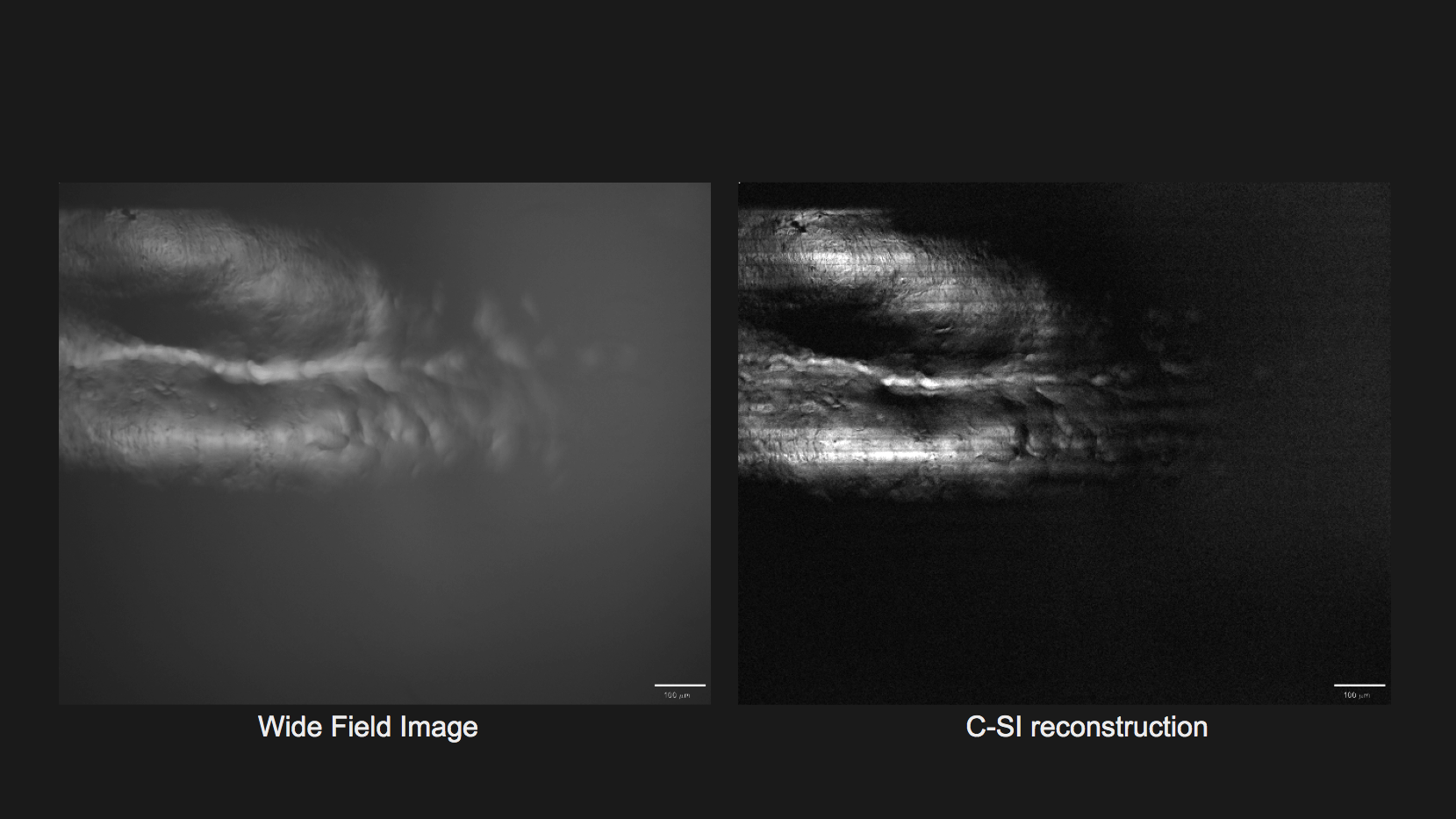
Structured Illumination for Low Scattering Imaging in Low-cost Microscopy
Micro-endoscopy is a GI cancer screening tool that can provide cellular resolution images in real-time. It works in conjunction with endoscopy and can reduce the need for biopsy. However, subsurface tissue scattering creates undesirable background on the image, reducing the contrast. While there are many systems such as point- scanning confocal and 2-photon microscopy that can reject scattering background, these system are typically very slow and expensive due to the optomechanical parts. We are proposing a structured illumination microscopy that is able to reject scattering while acquiring images in real time. We test background rejection ability of our system both theoretically and experimentally and show that it provides low scattering images while costing a fraction of the price of a traditional confocal system.
2017-2018
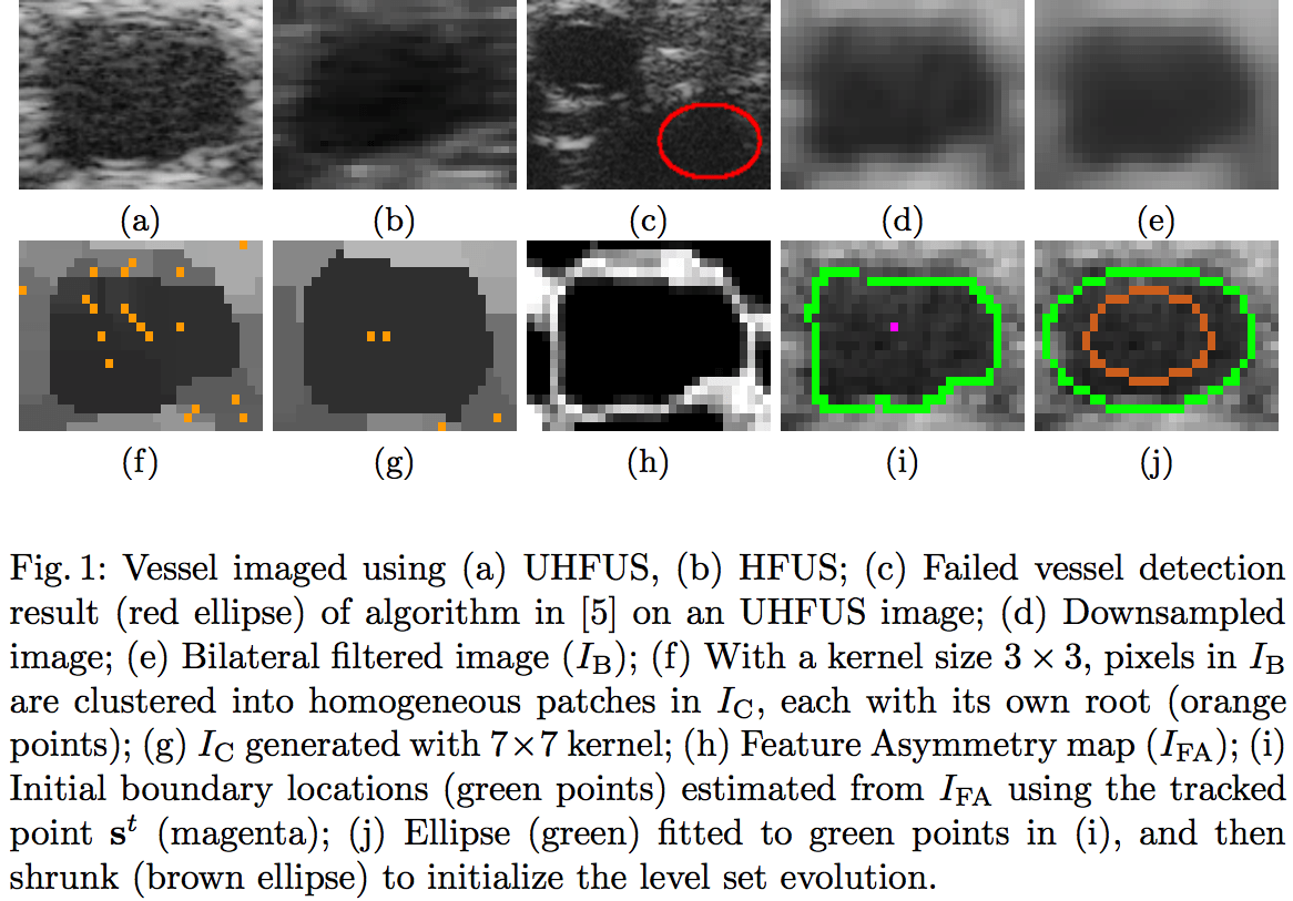
Vessel Tracking in Ultra-high Frequency Ultrasound Imaging
Ultra High-Frequency Ultrasound (UHFUS) enables the visualization of highly deformable small and medium vessels in the hand. Intricate vessel-based measurements, such as intimal wall thickness and vessel wall compliance, require sub-millimeter vessel tracking between B-scans. Our fast GPU-based approach combines the advantages of local phase analysis, a distance-regularized level set, and an Extended Kalman Filter (EKF), to rapidly segment and track the deforming vessel contour. We validated on 35 UHFUS sequences of vessels in the hand, and we show the transferability of the approach to 5 more diverse datasets acquired by a traditional High-Frequency Ultrasound (HFUS) machine. To the best of our knowledge, this is the first algorithm capable of rapidly segmenting and tracking deformable vessel contours in 2D UHFUS images. It is also the fastest and most accurate system for 2D HFUS images.
Tejas Sudharshan Mathai, Lingbo Jin, Vijay Gorantla, John M. Galeotti:
Fast Vessel Segmentation and Tracking in Ultra High-Frequency Ultrasound Images. MICCAI (4) 2018: 746-754
Awards
Rice University
President’s Prize
Fall 2018
Rice University
John Clark, Jr. Fellowship Award
Fall 2018
Carnegie Mellon University
Summer Undergraduate Research Fellowship
Summer 2017
American Heart Association
Carnegie Heart Fellowship
Summer 2016